
Mayfair • January 21, 2022
Having a small amount of fat in your liver is normal and may not cause any problems or symptoms. However, sometimes when the fat starts to build up it can be a cause for concern. In some cases, fat in the liver can cause it to become inflamed. This can damage your liver and cause scarring.
Liver fibrosis results from scarring and stiffening of healthy liver tissue. This form of liver damage can result from a variety of causes.
Liver damage can be caused by a variety of factors, including excessive alcohol use, fatty liver, or viral and autoimmune causes. Non-alcoholic fatty liver disease (NAFLD) is one of the most common types of chronic liver disease in Canada. It occurs when excess fat is stored in liver cells for no known reason and can lead to liver inflammation and ultimately liver fibrosis.
Note that not all patients with fatty liver have NAFLD. However, according to the Canadian Liver Foundation , NAFLD affects up to 20% of people in Canada. It often develops in people who are overweight or obese, or in people who eat a diet high in fat and sugar.
Your liver is the second largest organ in your body. It helps process nutrients and filters harmful substances from your blood. When a high fat buildup leads to liver fibrosis, it can prevent your liver from functioning properly.
As liver fibrosis progresses, the liver tissue becomes stiffer and can eventually lead to the final stage, referred to as cirrhosis.
Chronic liver disease can seriously affect liver function, but early diagnosis can limit further damage.
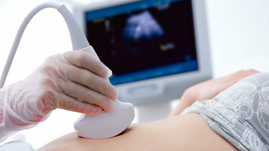
A type of ultrasound imaging called elastography directs painless, low-frequency vibrations at the liver to measure how quickly these vibrations move through it. A computer uses this information to create an optical map that shows the stiffness of the liver. This test is the same as an abdominal ultrasound with the use of elastography technology at the end of the test.
By measuring the stiffness of your liver, elastography is a suitable tool for looking for the variety of diseases that can occur in the liver and:
Traditionally, liver fibrosis was assessed with a biopsy — a needle was used to take a small sample of your liver tissue for examination under a microscope. However, this invasive technique can be associated with complications and only a small portion of the liver can be sampled.
With mild to moderate liver fibrosis, many people do not experience symptoms. Therefore, it can be difficult to diagnose chronic liver disease. Your doctor may look at risk factors, such as heavy alcohol use, obesity, and diabetes, to help make a diagnosis, as well as blood test results.
Ultrasound helps healthcare professionals make a diagnosis and inform care decisions. Once your doctor determines the need for this test, your doctor’s office can schedule an appointment for you or give you a number to call to schedule your appointment. You will also be given a request form and instructions for preparing for your test.
For a liver elastography ultrasound, you will be asked to fast and not have anything to eat or drink (except water) for six hours before your exam. This exam generally takes 20-30 minutes to complete.
Once you enter the exam room, you may be asked to change into a gown. You will then be positioned by one of our compassionate and experienced sonographers. A warm, fragrance-free, hypoallergenic, water-based ultrasound gel will be applied to your abdomen, and your sonographer will move the transducer around the front and sides of your abdomen and sides to gather images of your organs. You may be asked to hold your breath and change positions to help better examine the area of concern. You may experience mild to moderate pressure while the sonographer takes the images.
Your images will be reviewed by a qualified radiologist, who will prepare a report that will be sent to your doctor within 24 hours, earlier for urgent requests. Mayfair Diagnostics is owned and operated by over 50 radiologists, who are fellowship trained in many key areas such as neuroradiology, body, cardiac, musculoskeletal, etc. This allows for specialized review of your imaging by the appropriately trained radiologist.
Your images will be uploaded to a provincial Picture Archiving and Communication System (PACS) – this technology provides electronic storage and easy access to your medical images from many sources, including your doctor, specialists, hospitals and clinics.
Your doctor will review your images and the report from the radiologist and discuss next steps with you, such as a treatment plan or the need for further diagnostic imaging or laboratory tests to ensure an accurate diagnosis.
Our elastography services are available at our Castleridge , Coventry Hills, Mahogany Village and Market Mall locations in Calgary and our Queens location in Saskatchewan. For more information about our clinic locations and services, please visit our clinic location pages .
Canadian Liver Foundation (2022) “Fatty Liver Disease.” www.liver.ca. Accessed January 11, 2022.
Gherlan, GS (2015) “Ultrasound elastography of the liver: More than disease staging.” World Journal of Hepatology. Accessed January 11, 2022.
Nall, R. (2022) “Liver Fibrosis.” www.healthline.com. Accessed January 11, 2022.
Radiological Society of North America (2022) “Elastography.” www.radiologyinfo.org. Accessed January 11, 2022.